The idea that you are not alone inside your head sounds like a sci-fi plot twist. But recent data suggests your mother is, quite literally, part of your architecture.
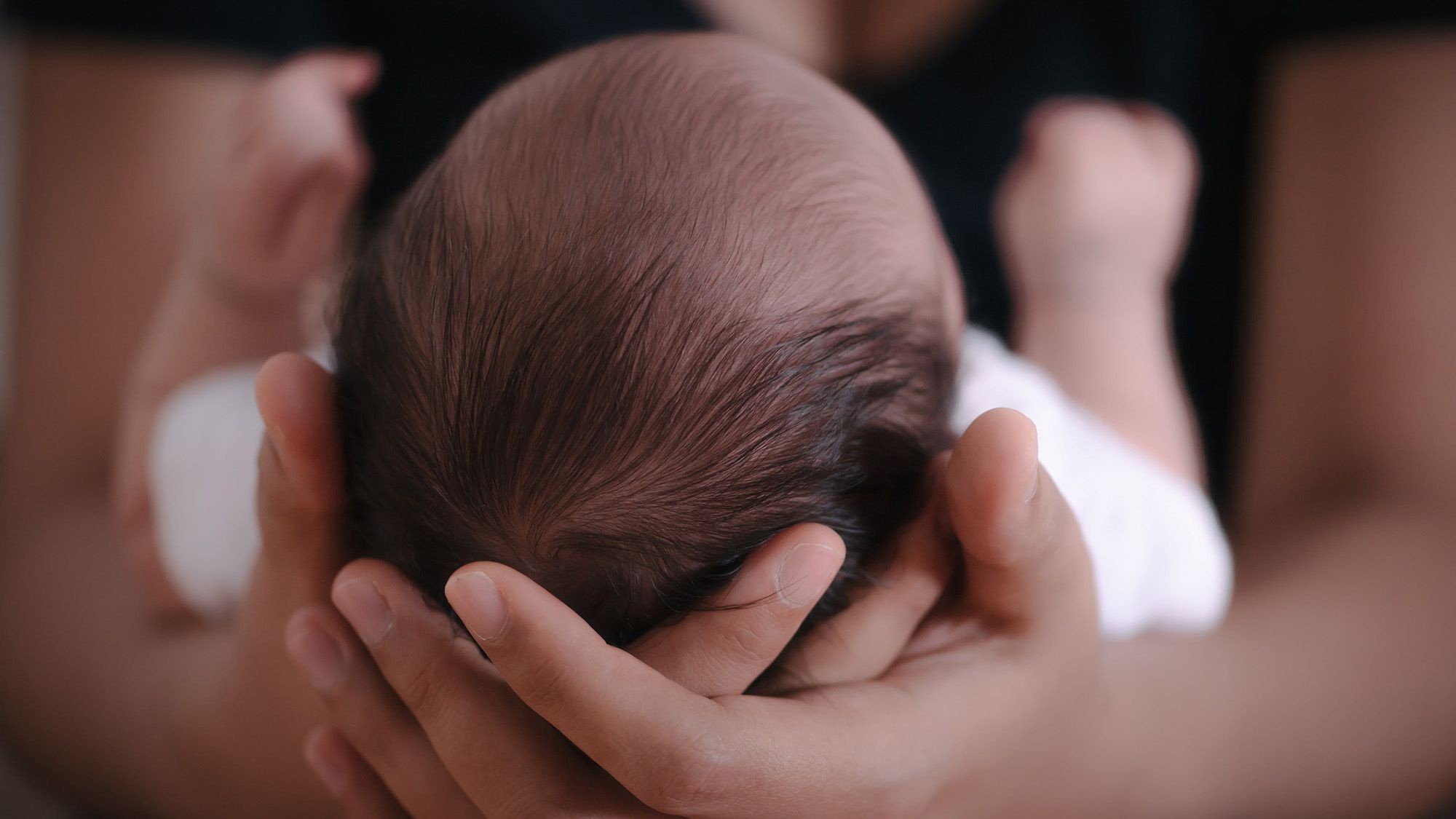
Scientists have confirmed that fetal brains contain cells bearing the mother’s DNA. These aren’t fleeting visitors. They stick around. They integrate. And they can persist into old age.
The research, published to the preprint server bioRxiv on June 10, adds weight to the phenomenon of microchimerism. It is the exchange of cells between a mother and her developing fetus during pregnancy. For years, we knew moms passed cells to kids. We knew the brain could harbor them. What was missing was a clear, tissue-level map of where they hide and how they function.
Previous studies relied on blood samples. Blood is a moving target, rich with circulating cells but poor at revealing what’s happening inside specific structures. This study looks at the tissue itself. Real human tissue. From real humans who had no say in the matter other than having existed.
The search for foreign DNA in fetal tissue
Getting brain samples from living children is ethically tricky. So the lead researcher, Sami Kanaan from Seattle’s Fred Hutchinson Cancer Center, took a pragmatic route. He analyzed brain tissue removed from dozens of young people undergoing surgery for severe epilepsy. These were not optional procedures. These were life-altering surgeries for children ranging from 28 days old to 19.
Kanaan’s team matched this tissue with cheek swab DNA from the mothers.
They used quantitative PCR. A sensitive tool designed to find needles in a haystack of billions of cells.
The results were stark. In 37 pairs of mother and child, 26 kids (70%) showed traces of maternal DNA.
It wasn’t a scattered dusting. It was systemic.
Maternal cells appeared in the frontal lobes. The temporal lobes. The parietal lobes. Even the hippocampus, the deep, wrinkled structure responsible for memory and emotion. On average, there were 2.2 maternal cells for every 100,001 cells. Standard numbers sound low until you do the math. One sample had 459 such cells. A significant number.
Importantly, not everyone carried them. 11 children had zero evidence. Why some had them and others didn’t is still up for debate. Being a firstborn might increase the odds. The data skewed that way. Fourteen of the kids with maternal cells were firstborns. Only one of those without them was a firstborn. But the sample size is too small to scream “conclusion.”
From bystanders to workers
Finding the cells was one thing. Knowing what they were doing was another.
Kanaan’s team employed single nucleus RNA sequencing. A technique that peeks into the cell to see which genes are switched on. It reveals identity. Purpose.
The maternal cells hadn’t just survived. They had adapted. They turned into working brain components.
Likely arriving as leukocytes or stem cells, these intruders integrated into the brain’s workforce. The study found evidence of neurons. Cells that process information. They found oligodendrocytes. These cells wrap around neurons, creating the insulation sheaths that speed up signals. There were astrocytes, supporting functions and fueling the neural engines. Microglia, the resident immune cells, were there too. Endothelial cells lining the blood vessels also bore the maternal signature.
“We’ve been so limited in understanding function,” noted Amy Boddy of UCSB. She was not part of this study. Her reaction? “This is amazing.”
Until now, animal models were the only guide. Animal brains are convenient. Human brains are messy. They are individual. This data comes from actual people.
Do they exist in healthy adults?
Epilepsy might skew the picture. Could the surgery, the trauma, or the neurological condition be drawing these cells in?
To test that, Kanaan and colleagues looked elsewhere.
They scanned autopsy data from 32 people with no neurodevelopmental disorders. Ages varied wildly. From 22 weeks gestation to 40-years-old. They also included brain tissue from three octogenarian/nonagenarians collected for an Alzheimer’s study. These men were in their 80s and 90s, free of known brain disease at the time of collection.
The result? Foreign cells appeared in 78% of these subjects too. Including a man in his 90ies.
Because the researchers lacked the mothers’ DNA for these healthy donors, they couldn’t definitively label them “maternal.” The cells could come from a twin. A previous pregnancy lost to miscarriage. An older sibling. A grandmother. But the likelihood leans heavily toward the mother. The source doesn’t matter less to the brain’s current state. They behave the same way.
They mature.
In younger brains, these foreign cells mostly became a specific type called a layer 2/3 neurons. In older brains? Microglia.
So the cells age with you. They switch jobs.
Are they useful?
Why would a host tolerate foreign DNA in the central command center of the body? Evolution rarely keeps waste. Not in major structures.
Dr. Sing Sing Way at Cincinnati Children’s Hospital notes that the diversity of cell types is fascinating. “Do we maybe need [them] to help out?” Boddy asks.
If the brain is constantly replacing or maintaining its network, having a backup supply of differentiated cells could be a survival mechanism. Or it could be a benign byproduct of being a mammal with a placenta. A side effect of intimacy between two genomes.
The number of these cells does drop as people age. They do not stay constant. But they don’t disappear, either.
Boddy suspects they are doing something vital. Or something complex. If we understand how microchimerism affects healthy brain development, we might unlock new avenues for understanding neurological disorders. Not just in the young. In the elderly, where the microglia dominate these foreign populations.
The limits of current tech
The study is robust. The methods are cutting-edge. But it’s not the final word.
Future research needs larger datasets. Uniform samples. The current work relies on surgical tissue (unavailable in healthy controls) and autopsies (often limited by cause of death or storage). To get clear answers, scientists need more biopsies. More cells analyzed per sample. Specimens gathered across specific ages to track decline. Identical brain regions mapped across different people.
Way points out that while the findings “push the boundaries,” we are still piecing together the puzzle with only the center images visible.
For now, we know this: your brain is a mosaic. It contains your genetic code. It contains traces of your parents. Your mother is in there. Somewhere. Probably working. Maybe remembering.
Who needs a therapist when you have your mom living in your temporal lobe?
It raises as many questions as it answers. Does the presence of these cells alter who you are? Do they change how your brain heals? Are you ever truly just you?
We may have to wait for better data to find out.